Physical and Chemical Responses in Animals
In 1924, Thomas Lewis and Ronald Grant at University College London injected histamine intradermally and recorded the 'triple response': a red flare from vasodilation, a wheal from increased capillary permeability, and a spreading flare from axon reflexes. Their experiment documented three of the four cardinal signs of inflammation at the cellular level, building on Henry Dale and Patrick Laidlaw's 1910 isolation of histamine from ergot. These five mediator classes are now the foundation of every anti-inflammatory drug in use today.
Orient yourself
Warm up on earlier material, predict what causes each sign of inflammation, then scan the goals and vocabulary.
Three quick questions from earlier lessons. Pulling old material back to mind before you learn something new makes the new material stick better, so this is not busywork.
Practise this lesson
Four printable worksheets that build from the foundations up to exam-style questions, start at whatever level suits you.
When you get a cut that becomes infected, the area around it turns red, swells, feels warm, and hurts. This is inflammation.
Before reading: predict what is actually happening at the cellular level to cause each of these four signs, redness, swelling, warmth, and pain. Write your prediction for each one.
Know
- Physical barriers that prevent pathogen entry in animals
- The four cardinal signs of inflammation and their causes
- The role of fever as a chemical defence
- Key chemical mediators: histamine, cytokines, complement
Understand
- Why inflammation is an adaptive response, not just a symptom
- How physical and chemical responses work together
- Why fever is beneficial within a range but dangerous if excessive
Can Do
- Link each sign of inflammation to its cellular cause
- Explain the sequence of events in the inflammatory response
- Analyse data showing physical and chemical changes in infected tissue
The castle walls
Map the barriers that stop most pathogens before any immune cell is involved, then sort them into physical and chemical.
Core Content
The body's castle walls
Right now, your skin is shedding approximately one million dead cells per hour, each removal carrying surface microbes with it. Your respiratory tract's cilia beat 1,000 times per minute to sweep a continuous mucus escalator toward your throat. Your stomach maintains a pH of 1.5–3.5, hostile to most bacteria. Most pathogens never get past these structural barriers. The immune response only activates when they do.
| Barrier | Location | How It Prevents Infection |
|---|---|---|
| Skin (epidermis) | External body surface | Tough, keratinised, multilayered, physically blocks most pathogens; dead outer cells constantly shed, removing surface microbes; slightly acidic pH inhibits bacterial growth |
| Mucous membranes | Respiratory, digestive, urogenital tracts | Mucus traps pathogens and particles; cilia sweep mucus toward exits (mucociliary escalator); goblet cells continuously replenish mucus layer |
| Cilia | Respiratory tract lining | Coordinated beating moves mucus and trapped pathogens upward toward throat for swallowing or expulsion |
| Stomach acid | Stomach | HCl (pH 1.5–3.5) destroys most ingested pathogens before they reach the intestine |
| Lysozyme | Tears, saliva, nasal secretions | Enzyme that degrades peptidoglycan in bacterial cell walls, bactericidal in mucosal secretions |
| Normal microbiome | Skin, gut, vaginal tract | Commensal bacteria compete with pathogens for nutrients and attachment sites; some produce antimicrobial compounds |
| Sebaceous glands | Skin | Secrete sebum, slightly acidic oil that creates an inhospitable environment for many pathogens on skin surface |
First-line (non-specific) barriers prevent entry before any immune response: skin (keratinised, acidic, sheds outer cells); mucous membranes + cilia trapping and sweeping pathogens (the mucociliary escalator); chemical barriers within them, stomach acid, lysozyme (degrades bacterial cell walls), and sebum; and the normal microbiome, which out-competes pathogens. The physical barrier is the most important defence, everything else is a fallback.
Pause, copy the main first-line barriers and how each blocks pathogens into your book.
Which of the following is a physical (structural) barrier to infection?
Four signs, one purpose
Trace each visible sign of inflammation back to the cellular change that causes it.
Redness · swelling · heat · pain
We just saw the barriers that keep pathogens out. That raises a question: what happens the moment a pathogen actually breaches them and enters tissue? This card answers it → the inflammatory response and its four cardinal signs.
The four cardinal signs of inflammation are not random symptoms, each has a specific cellular cause and a specific defensive purpose.
When a pathogen breaches physical barriers and enters tissue, the inflammatory response begins within seconds. Its purpose is to deliver immune cells and chemical mediators to the site of infection, contain the pathogen, and begin repair.

The four cardinal signs of inflammation, each caused by a specific cellular mechanism, each serving a defensive purpose
Four cardinal signs of inflammation, each with a cause: redness, vasodilation (more blood flow); swelling, increased capillary permeability (plasma leaks into tissue); heat, increased blood flow + immune-cell metabolism; pain, prostaglandins and bradykinin stimulate nociceptors. The purpose is to deliver immune cells and mediators to the infection site, contain the pathogen, and start repair.
Pause, copy the four signs with the cause of each into your book.
Redness and warmth at an infection site are both caused mainly by:
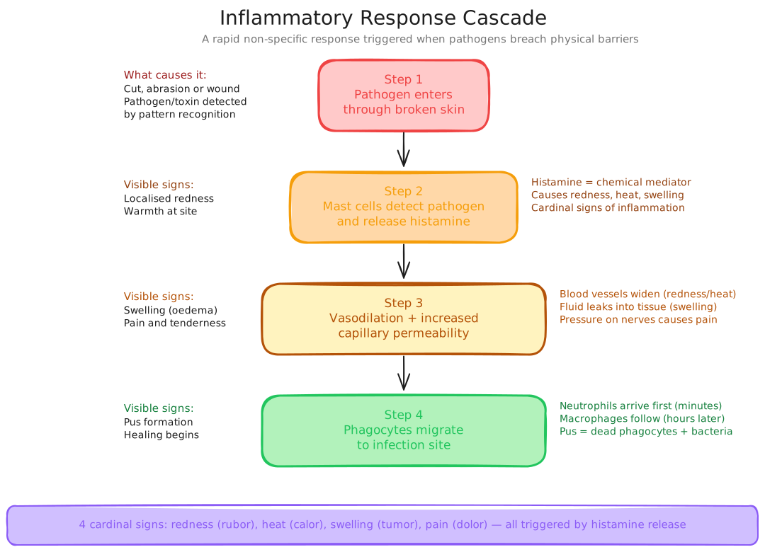
Inflammatory Response Cascade
The chemical chain of command
Follow the cascade in order, then learn which molecule is responsible for each step.
A precise sequence triggered by chemical signals
We just saw the four signs of inflammation. That raises a question: in what order do they actually arise, and what triggers each step? This card answers it → the inflammatory cascade from pathogen entry to pathogen destruction.
Inflammation does not just happen, it unfolds in a precise sequence triggered by chemical signals from damaged and infected cells.

The inflammatory cascade, from pathogen entry to immune cell recruitment and pathogen destruction
Inflammatory cascade sequence: pathogen enters → mast cells release histamine → vasodilation + increased capillary permeability → neutrophils migrate (drawn by a chemokine gradient) → pathogen destruction and tissue repair. Pus = dead neutrophils + pathogen debris, a sign the immune response actively engaged.
Pause, copy the inflammatory cascade sequence in order into your book.
Mast cells release _____, which causes vasodilation and increased capillary permeability at the infection site.
The signalling molecules that coordinate inflammation
We just saw the cascade naming histamine, chemokines and neutrophils. That raises a question: what are all the signalling molecules behind it, and what does each one do? This card answers it → the chemical mediators that coordinate the whole inflammatory response.
The inflammatory response is coordinated by chemical signals, understanding what each does is essential for the HSC.
Histamine
Prostaglandins
Cytokines
Chemokines
Complement proteins
Interferons
Key chemical mediators: histamine, vasodilation + permeability (from mast cells/basophils); prostaglandins, pain + fever; cytokines (IL-1, IL-6, TNF-α), recruit immune cells and trigger fever; chemokines, attract phagocytes along a gradient; complement, opsonisation + membrane attack complex; interferons, antiviral signal warning neighbouring cells.
Pause, copy each mediator and its one-line role into your book.
Which chemical mediator signals neighbouring cells to produce antiviral proteins?
Fever on trial
Treat fever as a deliberate response, then judge with real data whether suppressing it helps or harms.
A deliberate response coordinated by the hypothalamus
We just saw that cytokines and prostaglandins act as pyrogens. That raises a question: what whole-body response do those pyrogens trigger? This card answers it → fever, a systemic, hypothalamus-controlled defence.
Fever is not just an unfortunate side effect of infection, it is a deliberate, adaptive response coordinated by the hypothalamus.
The hypothalamus raises the body temperature set point in response to chemical signals, primarily pyrogens like IL-1 and prostaglandins, released by the immune system. The body then shivers and constricts surface blood vessels to reach the new, higher set point. A moderate temperature rise speeds up immune cell enzyme reactions and slows the replication of some pathogens, while a very high or prolonged fever (above ~40°C) risks denaturing the body's own proteins and requires treatment.
Fever: pyrogens (IL-1, prostaglandins, bacterial LPS) reach the hypothalamus, which resets the temperature "thermostat" upward → shivering and vasoconstriction raise body temperature. Benefit: speeds immune reactions and slows some pathogen replication. Danger: above ~40°C the body's own proteins risk denaturation.
Pause, copy how fever is generated and its benefit vs danger into your book.
A moderate fever can be beneficial because it speeds up immune reactions and slows some pathogen growth.
Lysozyme in tears and saliva destroys bacterial cell walls by breaking down peptidoglycan.
The skin is an ineffective physical barrier because pathogens can easily pass through dead keratinised cells.
Data Analysis, Temperature and Immune Function
Pattern A, Structured Data Analysis
The table below shows data from an experiment investigating the effect of temperature on neutrophil phagocytosis rate and bacterial replication rate in vitro (in cell culture).
| Temperature (°C) | Neutrophil phagocytosis rate (bacteria/neutrophil/hour) | Bacterial replication rate (doublings/hour) |
|---|---|---|
| 36.0 (sub-normal) | 4.1 | 2.8 |
| 37.0 (normal body temp) | 5.2 | 2.9 |
| 38.0 (mild fever) | 6.8 | 2.7 |
| 38.5 (moderate fever) | 7.4 | 2.4 |
| 39.5 (high fever) | 7.1 | 2.1 |
| 40.5 (very high fever) | 5.3 | 1.8 |
| 41.5 (dangerous fever) | 2.9 | 1.5 |
- Describe the trend in neutrophil phagocytosis rate as temperature increases from 37°C to 41.5°C.
- At what temperature does neutrophil phagocytosis reach its peak? What happens to phagocytosis rate above this temperature, and suggest a biological explanation.
- Describe the trend in bacterial replication rate as temperature increases. How does this support the adaptive value of fever?
- At 40.5°C, the neutrophil phagocytosis rate has declined from its peak but bacterial replication rate is also at its second lowest. Evaluate whether a fever of 40.5°C is likely to be beneficial or harmful to the host, using both columns of data.
- A doctor recommends treating any fever above 38°C with ibuprofen immediately. Evaluate this recommendation using the data provided.
When you get a splinter that becomes infected, a precise sequence unfolds. Bacteria enter the wound. Mast cells in the surrounding tissue release histamine, causing the familiar redness and warmth as blood vessels dilate and more blood arrives. Capillaries become leaky, plasma floods the tissue, causing the swelling. Prostaglandins sensitise nerve endings, causing the throbbing pain that tells you not to use the injured finger. Within hours, neutrophils arrive via chemokine gradients, engulf bacteria, and die, their remains accumulating as pus. Meanwhile, cytokines from macrophages at the site travel via the bloodstream to the hypothalamus, triggering a mild fever that accelerates the whole process. If the infection spreads, complement proteins in the blood coat bacteria for easier phagocytosis. Each of these steps, once dismissed as mere "symptoms", is a precisely coordinated defence mechanism. The redness, swelling, heat, and pain of a small infected cut represent your body running a 400-million-year-old programme that is extraordinarily effective at what it does. You will map this process in the practice questions.
Physical Barriers
- Skin, keratinised, acidic, constantly shedding.
- Mucous membranes + cilia, trap and sweep pathogens.
- Stomach acid (pH 1.5–3.5), destroys ingested pathogens.
- Lysozyme in tears/saliva, degrades bacterial cell walls.
Four Signs of Inflammation
- Redness, vasodilation → more blood flow.
- Swelling, increased capillary permeability → plasma leaks into tissue.
- Heat, increased blood flow + immune cell metabolism.
- Pain, prostaglandins + bradykinin stimulate nociceptors.
Key Chemical Mediators
- Histamine: vasodilation + permeability (mast cells).
- Prostaglandins: pain + fever.
- Cytokines (IL-1, IL-6, TNF-α): recruit immune cells; trigger fever.
- Complement: opsonisation + membrane attack complex.
- Interferons: antiviral, signal neighbouring cells.
Fever
- Trigger: pyrogens (IL-1, prostaglandins, bacterial LPS) reach hypothalamus.
- Mechanism: hypothalamus resets thermostat upward → shivering/vasoconstriction.
- Benefit: speeds immune reactions; reduces some pathogen replication.
- Dangerous above 40°C, protein denaturation risk.

Inflammatory Response, Step by Step
Practise independently
Attempt at least one response in your own words. Model answers support checking; they do not replace the attempt.
A fresh set drawn from this lesson's question bank, feedback shown immediately. +5 XP per correct · +25 XP all correct
Pick your answer, then rate your confidence, that tells the system what to drill next.
UnderstandBand 3(3 marks) 1. Explain how the skin and mucous membranes act as physical barriers against pathogen entry. For each, identify one structural feature and explain how it prevents infection.
1 mark: skin feature + mechanism · 1 mark: mucous membrane feature + mechanism · 1 mark: one additional feature (lysozyme, cilia, stomach acid, microbiome)
EvaluateBand 4(3 marks) 2. A student claims: "Inflammation is a harmful overreaction by the immune system, anti-inflammatory drugs should always be taken to reduce it." Evaluate this claim, referring to the causes and purposes of the four cardinal signs of inflammation.
1 mark: identifies what causes the four signs · 1 mark: explains the defensive purpose of at least two signs · 1 mark: evaluative conclusion
AnalyseBand 5(4 marks) 3. Describe the sequence of physical and chemical changes that occur in host animal tissue in the first 12 hours following bacterial infection of a skin wound. In your answer, refer to at least four specific mediators or cell types and explain the role of each.
1 mark per correctly described mediator/cell type with its role (max 4): mast cells/histamine, prostaglandins, cytokines/chemokines, neutrophils, complement, interferons
Show all answers
Multiple choice
MC answers and full explanations are shown inline as you complete each question. Use the retry button to attempt a fresh set from the lesson bank.
Short Answer Model Answers
Q1 (3 marks): The skin acts as a physical barrier through its keratinised, multilayered epidermis, keratin makes the outer skin tough and relatively impermeable, and the constant shedding of dead outer cells removes any surface-dwelling microorganisms before they can establish infection. The skin's slightly acidic pH (around 4.5–5.5) also inhibits the growth of many pathogenic bacteria. Mucous membranes line the respiratory, digestive, and urogenital tracts. Mucus physically traps pathogens in a sticky gel layer, and the coordinated beating of cilia sweeps this mucus (with trapped pathogens) upward toward the throat in the mucociliary escalator, where it is swallowed and destroyed by stomach acid. An additional physical defence is lysozyme, present in tears, saliva, and nasal secretions, this enzyme degrades peptidoglycan in bacterial cell walls, killing bacteria on contact at mucosal surfaces.
Q2 (3 marks): The student's claim is incorrect. The four cardinal signs of inflammation each serve defined defensive purposes. Redness and heat are caused by vasodilation (triggered by histamine), increasing blood flow to deliver immune cells and chemical mediators rapidly. Swelling is caused by increased capillary permeability, allowing plasma (including antibodies and complement proteins) to flood the infection site. Pain (from prostaglandins and bradykinin) signals tissue damage and reduces use of the injured area, protecting it during repair. These responses are adaptive mechanisms essential for pathogen clearance and tissue repair. Anti-inflammatory drugs are appropriate when inflammation is excessive, prolonged, or causing more harm than benefit, but taking them reflexively suppresses responses that are actively assisting recovery and may prolong infection clearance. The claim is an oversimplification.
Q3 (4 marks): Within seconds of bacterial entry, damaged tissue cells and mast cells release histamine, causing vasodilation (increasing blood flow → redness and warmth) and increased capillary permeability (allowing plasma to leak into tissue → swelling), and delivering complement proteins to the site. Simultaneously, damaged cells release prostaglandins and bradykinin, sensitising pain receptors (nociceptors) → pain. Within the first hour, macrophages phagocytose bacteria and release cytokines (IL-1, IL-6, TNF-α) which travel to the hypothalamus triggering fever, and act as chemokines establishing a gradient that attracts neutrophils (chemotaxis). Within 2–6 hours, neutrophils migrate out of blood vessels (diapedesis) and engulf bacteria by phagocytosis, killing them with reactive oxygen species; dead neutrophils accumulate as pus. Complement proteins coat bacteria (opsonisation) and form membrane attack complexes that puncture bacterial membranes. By 12 hours, this combination of physical, cellular, and chemical responses has typically contained the infection and begun repair.
Retrieve and reflect
Use the Review session first, then compare how your thinking changed.
A full module quiz covering every lesson in this module, not just this one. Set aside a decent block of time and treat it like a real assessment.
Start the module quiz →You were asked to predict the cellular cause of each of the four signs of inflammation, redness, swelling, warmth, and pain. Thomas Lewis and Ronald Grant's 1924 UCL experiments confirmed the mechanisms for three of them with a single reagent: intradermal histamine injection produced a red flare (vasodilation), a wheal (increased capillary permeability → oedema), and an outer spreading ring (axon reflex). Three observable signs, one chemical mediator.
The answers: redness and warmth share the same cause, vasodilation triggered by histamine from mast cells, which widens blood vessels and increases blood flow, bringing more warm blood close to the surface. Swelling has a different cause, increased capillary permeability (also triggered by histamine and other mediators) allows plasma to leak from blood vessels into the tissue space, causing fluid accumulation. Pain is caused by prostaglandins and bradykinin directly stimulating pain receptor nerve endings (nociceptors).
If you predicted that redness and swelling had the same cause, they are both triggered by histamine, but through different mechanisms (vasodilation vs permeability). If you predicted that warmth was caused by fever, local warmth at an infection site is due to increased blood flow, not systemic fever. Fever is a separate, whole-body response coordinated by the hypothalamus.
The key insight: every sign has a specific cellular mechanism and a specific defensive purpose. They are not random discomforts, they are information and action, happening simultaneously.