Adaptive Immunity, Antigens and Antibodies
In 1957, Frank Macfarlane Burnet at the Walter and Eliza Hall Institute in Melbourne proposed the clonal selection theory: the body maintains a library of B cells, each with a unique receptor, and infection selects the matching clone for expansion into an antibody-secreting army. A single activated B cell can produce up to 2,000 antibody molecules per second. Burnet's theory won the 1960 Nobel Prize in Physiology and is now the accepted mechanism of adaptive immunity.
Practise this lesson
Four printable worksheets that build from the foundations up to exam-style questions, start at whatever level suits you.
You had chickenpox as a child. Twenty years later, someone sneezes chickenpox virus near you. You don't get sick.
Before reading: at the molecular level, what do you think is preventing you from getting chickenpox a second time? Where is the "memory" stored, and how does it work fast enough to stop an infection that moves quickly?
Know
- What an antigen is and where antigens are found
- The structure and function of antibodies
- Clonal selection and clonal expansion
- The difference between plasma cells and memory B cells
Understand
- Why clonal selection is the key to specificity in adaptive immunity
- How antibodies neutralise pathogens through different mechanisms
- Why the secondary response is faster and stronger
Can Do
- Describe the sequence from antigen exposure to antibody production
- Explain clonal selection using the lock-and-key analogy
- Compare the primary and secondary immune responses using a graph
Core Content
Antigens and the epitopes antibodies recognise
In 1957, Frank Macfarlane Burnet's clonal selection theory proposed that B cells carry receptors matching specific molecular features on foreign particles. The surface spike protein of the SARS-CoV-2 virus carries hundreds of different molecular shapes, each one a potential antigen. An antibody raised against the spike protein binds to just one small region of it: the epitope. That single binding event is enough to block the virus from entering a cell.
An antigen is any molecule recognised by the adaptive immune system that triggers a specific immune response. Most antigens are proteins or polysaccharides found on the surface of pathogens, but they can also be found on pollen, transplanted cells, or even the body's own abnormal cells in autoimmune disease.
The part of the antigen that is actually recognised by an antibody or lymphocyte receptor is called the epitope (or antigenic determinant). An epitope is the specific region of an antigen that an antibody binds to, and one antigen can have many epitopes, each stimulating a different B cell clone.
Pause, copy the highlighted points into your book before moving on.
The immune system tolerates self-antigens (your own normal molecules) but attacks non-self antigens (foreign molecules), this self vs non-self recognition is the basis of immune specificity.
Add the self vs non-self point to your notes before the check below.
The specific part of an antigen that an antibody actually binds to is called the _____.
A Y-shaped protein with a unique binding site
We just saw that an antigen carries many small epitopes. That raises a question: what is it about an antibody that lets it lock onto just one of them? This card answers it → the antibody's Y-shape with its one unique variable binding site.
Every antibody has the same basic Y-shape, but a unique antigen-binding site that matches exactly one epitope.
Antibodies (immunoglobulins) are Y-shaped proteins made by plasma cells. The variable region is the unique antigen-binding site (fits one epitope); the constant (Fc) region sets the antibody's class and effector functions. Each antibody has two identical binding sites.
Antibody structure, the variable regions give each antibody its unique specificity; the constant region determines its class and effector functions
Pause, copy the highlighted structure points, using the diagram to label variable vs constant regions.
Antibodies neutralise pathogens through several mechanisms:
- Neutralisation: antibody binds to pathogen surface, physically blocking it from attaching to host cell receptors
- Opsonisation: antibody coating marks pathogen for phagocytosis, phagocytes have Fc receptors that bind the constant region
- Complement activation: antibody-antigen complexes activate the complement cascade, leading to membrane attack complex formation
- Agglutination: antibodies clump pathogens together (each antibody has two binding sites), making them easier to phagocytose and preventing spread
Four antibody actions: neutralisation (block attachment), opsonisation (mark for phagocytosis), complement activation (membrane attack complex), agglutination (clumping). Antibodies mark or block pathogens, they don't kill them directly.
Add the four antibody actions to your notes before the check below.
Which part of an antibody determines which specific antigen it can bind?
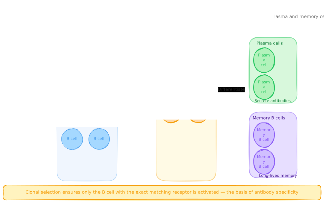
Clonal Selection and Expansion
Finding and multiplying the one matching B cell
We just saw that every antibody fits only one specific epitope. That raises a problem: the body can't pre-make an antibody for every possible germ. This card answers it → clonal selection is how the body finds and mass-produces the one matching B cell on demand.
The body holds millions of different B cell clones, each able to bind only one antigen, clonal selection is how the right one is found and turned into an army.
Each B cell has a unique B cell receptor (BCR) that binds only one specific antigen. Before any infection, most of these B cells are naive (never exposed to their antigen).
Clonal selection and expansion, one matching B cell becomes an army of plasma cells AND a bank of memory cells for future protection
Clonal selection: the antigen selects the one B cell whose BCR matches it. With a T helper co-stimulatory signal, that B cell undergoes clonal expansion, dividing into plasma cells (short-lived antibody factories) and memory B cells (long-lived, for a faster second response).
Pause, copy the highlighted clonal selection sequence, using the flowchart to get the order right.
During clonal selection, how many B cell clones are activated by a given antigen?
Annotated Diagram, Clonal Selection and Antibody Production
Pattern A, Draw and Annotate
In your book, draw a diagram showing the full sequence from antigen entry to antibody release. Your diagram must include and label:
- An antigen-presenting dendritic cell displaying antigen fragments on MHC II.
- A population of at least five different naive B cells with different-shaped BCRs, only one matches the antigen.
- The selected B cell binding the antigen and receiving a signal from a T helper cell.
- Clonal expansion, show the selected B cell dividing into multiple identical cells.
- Two final outcomes: a cluster of plasma cells releasing Y-shaped antibodies, and a separate cluster of memory B cells.
- An arrow showing antibodies binding to the original antigen, with a label explaining the effector function (choose one: neutralisation, opsonisation, or agglutination).
Why the second exposure is so much faster
We just saw that clonal selection leaves behind a reserve of memory B cells. That raises a question: what difference do those memory cells actually make next time? This card answers it → they explain why the second response is far faster and stronger than the first.
The dramatic difference between first and second exposure to an antigen is explained entirely by memory B cells.
The secondary response is faster (memory B cells activate within hours) and produces far more antibody, this is the basis of vaccination and lifelong immunity
Primary response: 7–14 days, lower antibody level, IgM then IgG. Secondary response: 1–3 days, much higher and longer-lasting level, mainly IgG, because memory B cells activate immediately. This is the basis of vaccination and lifelong immunity.
Pause, copy the highlighted primary vs secondary comparison, and sketch the graph shape.
The secondary immune response is faster and produces more antibody than the primary response.
Clonal selection activates only the B cell clone whose receptor matches a specific antigen.
Antibodies are produced by T cells and directly kill pathogens by piercing their cell membranes.
Interpreting Primary and Secondary Response Data
Pattern A, Structured Data Analysis
The table below shows antibody levels (arbitrary units) measured in a patient's blood following two exposures to the same pathogen.
| Day | Antibody level (AU) | Event |
|---|---|---|
| 0 | 0 | First exposure to pathogen |
| 5 | 2 | |
| 10 | 45 | |
| 14 | 80 | Peak primary response |
| 21 | 40 | |
| 35 | 12 | |
| 60 | 8 | Second exposure to same pathogen |
| 62 | 35 | |
| 64 | 180 | |
| 67 | 420 | Peak secondary response |
| 80 | 200 | |
| 100 | 95 |
- Draw a labelled graph of antibody level (y-axis) vs day (x-axis). Mark the two exposure events and label the primary and secondary response peaks.
- Calculate the ratio of peak secondary antibody level to peak primary antibody level. What does this ratio indicate about the effectiveness of immunological memory?
- The primary response took 14 days to reach its peak. The secondary response reached its peak by day 67, only 7 days after the second exposure. Explain this difference at the cellular level.
- After day 35, antibody levels in the primary response declined to 12 AU by day 60. Yet when the second exposure occurred, the patient responded rapidly. What does this suggest about where immunological memory is stored?
- A student claims: "The patient was immune after the first exposure because antibody levels didn't drop to zero." Evaluate this claim using the data.
When you were infected with the varicella-zoster virus (chickenpox) as a child, your adaptive immune system mounted a primary response: dendritic cells presented viral antigens, the matching B cell clone was selected and expanded, plasma cells flooded your bloodstream with anti-varicella antibodies, and memory B cells formed. The infection resolved in 1–2 weeks. The plasma cells died within weeks. But the memory B cells persisted, some for the rest of your life. Twenty years later, when the same virus enters your respiratory tract, those memory B cells are activated within hours. They divide rapidly into new plasma cells and flood the bloodstream with high-affinity IgG antibodies before the virus can establish a significant infection. You experience no symptoms, or only a very mild response, because the secondary immune response eliminates the virus before it reaches the threshold for illness. This is also why the chickenpox vaccine works: it introduces a weakened form of the virus that stimulates the same primary response and memory formation, without causing the disease. You will apply this to primary vs secondary response graphs in the practice questions.
Antigens and Antibodies
- Antigen: any foreign molecule triggering an immune response.
- Epitope: the specific region an antibody binds to.
- Antibody: Y-shaped protein; variable region = antigen-specific; Fc region = effector functions.
- Each antibody has TWO identical antigen-binding sites.
Clonal Selection and Expansion
- Millions of B cells, each with unique BCR, only one matches any given antigen.
- Matching B cell binds antigen + receives T helper signal → activated.
- Activated B cell divides → plasma cells (antibody factories) + memory B cells.
- T helper signal required, prevents accidental self-activation.
Antibody Functions
- Neutralisation, blocks pathogen from binding host receptors.
- Opsonisation, coats pathogen for phagocytosis.
- Complement activation, triggers membrane attack complex.
- Agglutination, clumps pathogens together.
Primary vs Secondary Response
- Primary: 7–14 days to peak; lower antibody levels; IgM then IgG.
- Secondary: 1–3 days to peak; much higher levels; mainly IgG.
- Secondary faster because memory B cells are pre-existing and activate immediately.
- Basis of vaccination, primary response without full disease.
B Cell Activation Pathway
A fresh set drawn from this lesson's question bank, feedback shown immediately. +5 XP per correct · +25 XP all correct
Pick your answer, then rate your confidence, that tells the system what to drill next.
UnderstandBand 3(3 marks) 1. Explain the process of clonal selection and clonal expansion. In your answer, describe what happens to the selected B cell and explain why the vast majority of B cells in the body are not activated during a typical infection.
1 mark: clonal selection (specific BCR matches antigen + T helper signal) · 1 mark: clonal expansion (rapid division into plasma cells and memory B cells) · 1 mark: why other B cells are unaffected (specificity)
ApplyBand 4(3 marks) 2. Describe two different mechanisms by which antibodies defend against pathogens. For each, explain what the antibody does and how this leads to pathogen elimination.
1 mark per mechanism correctly named and explained (max 2) · 1 mark: clear explanation of how each leads to elimination
EvaluateBand 5(4 marks) 3. Explain why a person who had chickenpox as a child is protected against chickenpox for life, but still needs an annual influenza vaccine. In your answer, refer to clonal selection, memory B cells, and the concept of antigen variation.
1 mark: chickenpox, memory B cells persist · 1 mark: secondary response mechanism · 1 mark: influenza mutates, new antigens · 1 mark: annual vaccine provides new primary response
Show all answers
Multiple choice
MC answers and full explanations are shown inline as you complete each question. Use the retry button to attempt a fresh set from the lesson bank.
Short Answer Model Answers
Q1 (3 marks): Clonal selection occurs when an antigen (presented on MHC II by a dendritic cell or macrophage) encounters the pool of naive B cells in a lymph node. Each B cell has a unique B cell receptor (BCR) that can bind only one specific antigen. The rare B cell whose BCR matches the antigen binds it and, crucially, receives a co-stimulatory signal from a T helper cell that has independently recognised the same antigen, this dual signal is required for full activation. The selected B cell then undergoes clonal expansion: it divides rapidly and repeatedly, producing a large clone of genetically identical cells. These differentiate into two populations: plasma cells (which secrete thousands of identical antibodies per second for days to weeks) and memory B cells (long-lived cells that persist for years, ready for rapid re-activation). The vast majority of B cells in the body are unaffected because their BCRs do not match the specific antigen, only the exact matching clone is selected.
Q2 (3 marks): Neutralisation: antibodies bind to surface molecules on a pathogen, for example, the spike protein of a virus. By physically occupying the binding site, the antibody prevents the pathogen from attaching to its host cell receptor. A virus that cannot bind to a host cell cannot enter, replicate, or cause infection, so neutralisation prevents the infection spreading to new cells. Opsonisation: antibodies coat the surface of a pathogen (binding via their variable regions). Phagocytes have Fc receptors that bind the constant (Fc) region of antibodies, so an opsonised pathogen is held tightly to the phagocyte's surface, making adherence dramatically more efficient and leading to phagocytosis, the phagocyte engulfs the pathogen and lysosomal enzymes destroy it.
Q3 (4 marks): When a child is infected with varicella-zoster virus (chickenpox), the specific B cell clone with a matching BCR undergoes clonal selection and expansion, producing both plasma cells and memory B cells that persist in the lymph nodes and bone marrow, sometimes for life. On re-exposure (even decades later), these memory B cells activate within hours, rapidly dividing into plasma cells that flood the bloodstream with high-affinity IgG before the virus establishes a significant infection, so the person experiences no symptoms. Influenza requires annual vaccination because influenza A mutates rapidly, its surface antigens (haemagglutinin and neuraminidase) change from year to year through antigenic drift. Memory B cells formed against last year's strain carry receptors matched to last year's antigens; when this year's strain presents different antigens, those memory cells do not recognise them, so the new strain effectively requires a new primary response. Each annual vaccine introduces antigens from the strains predicted to circulate that season, generating new memory cells matched to those strains.
Five timed questions on antigens, antibodies, and adaptive immunity. Beat the boss to bank a tier, gold (perfect + fast), silver (80%+), or bronze (cleared).
⚔ Enter the arenaClimb platforms using your knowledge of antigens, antibodies and adaptive immune responses. Pool: lessons 1–11.
You were asked where chickenpox immunity is stored and how it works fast enough to stop a rapidly moving infection. Frank Macfarlane Burnet's 1957 clonal selection theory, for which he received the 1960 Nobel Prize, gives the answer: the immune system maintains a library of B cells, each with a unique receptor, and infection selects the matching clone. That clone produces not only short-lived plasma cells but long-lived memory B cells that persist in lymph nodes and bone marrow for years or decades.
The memory is stored in memory B cells, long-lived lymphocytes that formed during the primary response. They carry exactly the same BCR as the original selected B cell, meaning they recognise the same varicella-zoster antigens.
They work fast enough because they do not need to go through the slow process of clonal selection from a naive pool. They are already selected, already matched, already present in much larger numbers than the original naive clone. On re-exposure, they activate and begin dividing within hours, producing plasma cells that release antibodies within 1–3 days. The virus is cleared before it can establish enough of an infection to cause symptoms.
If you predicted "antibodies are already in the blood", that is partially right for shortly after infection, but antibody levels do decline over months to years. The key insight is that the memory is cellular (stored in long-lived B cells), not just chemical (stored as pre-existing antibodies). Antibodies are re-made on demand by memory cells reactivating, they are not simply stockpiled indefinitely.