The Innate Immune System
On 14 December 1882, Élie Metchnikoff inserted a rose thorn into a transparent starfish larva and watched through his microscope. Within hours, mobile cells had surrounded and engulfed the thorn. He described phagocytosis, the cellular engulfment of foreign material, in Virchows Archiv in 1883, and shared the 1908 Nobel Prize in Physiology with Paul Ehrlich. The neutrophils, macrophages, and dendritic cells he discovered are now understood as the core cellular machinery of the innate immune system.
Practise this lesson
Four printable worksheets that build from the foundations up to exam-style questions, start at whatever level suits you.
You get a splinter in your finger at 9am. By 9:15am the area is red and starting to swell, before you've even thought about cleaning it.
Before reading: predict the sequence of events happening in that 15-minute window. What cells or molecules do you think are involved, and in what order do they act? Be as specific as you can.
Know
- The components of the innate immune system
- How phagocytosis works, step by step
- The role of natural killer cells
- How the innate system differs from the adaptive system
Understand
- Why the innate response is fast but non-specific
- How pattern recognition allows innate cells to detect pathogens
- Why NK cells target infected host cells rather than pathogens directly
Can Do
- Describe phagocytosis as a sequence of steps
- Distinguish innate from adaptive immunity with examples
- Apply innate immune components to an unfamiliar infection scenario
Core Content
Fast and broad vs slow and specific
A splinter enters your fingertip at 9:00 AM. By 9:01, mast cells at the injury site have already released histamine, blood vessels are dilating and permeating. By 9:15, the first neutrophils have arrived and begun engulfing bacteria. No antibodies. No prior exposure to that bacterium needed. This immediate, automatic response is the innate immune system, and it was running before you even noticed the splinter.
Two integrated layers. Innate: fast (minutes), non-specific, no memory; PRRs detect PAMPs; cells = neutrophils, macrophages, dendritic cells, NK cells, mast cells. Adaptive: slow (days), highly specific, has immunological memory; B and T lymphocytes with antigen receptors. The two layers work together, dendritic cells bridge them by presenting antigens to T cells.
Pause, copy the innate vs adaptive comparison (speed, specificity, memory, cells) into your book.
Compared to adaptive immunity, the innate immune response is:
Apply to an Unfamiliar Organism, The Sea Urchin
Pattern C, Apply to Unfamiliar Context
Sea urchins (Strongylocentrotus purpuratus) have been extensively studied as a model organism for innate immunity. They have no adaptive immune system, no B cells, no T cells, no antibodies. Yet they survive in bacteria-rich marine environments and can live for decades. Their immune system consists entirely of innate mechanisms, and their genome contains over 200 different pattern recognition receptor genes, far more than humans.
- Sea urchins have no adaptive immune system. Predict two ways this limits their ability to respond to pathogens compared to vertebrates.
- Sea urchins have over 200 pattern recognition receptor genes. Explain why a larger repertoire of PRRs might compensate for the absence of an adaptive immune system.
- A sea urchin is injected with Vibrio bacteria. Predict which innate immune responses, using only mechanisms described in this lesson, would be activated. Be specific about which cells or molecules would be involved.
- Sea urchins can live for over 100 years despite having no adaptive immunity. What does this suggest about the relative effectiveness of the innate immune system in environments where pathogen diversity is limited and stable?
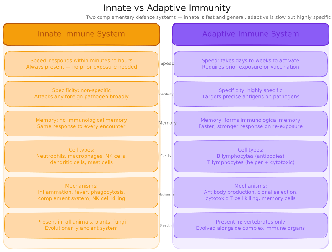
Innate vs Adaptive Immunity Comparison
Cellular and soluble components
We just saw that the innate system is the fast, non-specific first layer. That raises a question: what actually makes it up? This card answers it → the cellular and soluble components of innate immunity.
The innate immune system has three layers: physical barriers (L09), cellular components, and soluble chemical components, this lesson focuses on the cellular response.
| Component | Type | Function |
|---|---|---|
| Neutrophils | White blood cell (first responder) | First phagocytes to arrive at infection site; engulf and destroy bacteria; short-lived (die within hours-days); most abundant WBC in blood |
| Macrophages | White blood cell (tissue resident) | Long-lived phagocytes patrolling tissues; engulf pathogens and debris; release cytokines; present antigens to T cells (bridge to adaptive) |
| Dendritic cells | White blood cell (sentinel) | Patrol tissues for pathogens; engulf and process antigens; migrate to lymph nodes to activate T cells, key link between innate and adaptive |
| Natural killer (NK) cells | Innate lymphocyte | Target and kill virus-infected host cells and cancer cells; do not need antigen presentation, detect absence of MHC I markers |
| Mast cells | Tissue resident cell | Release histamine and other mediators on pathogen detection; initiate inflammation; important in allergy responses |
| Complement system | Soluble proteins (in blood) | Coat pathogens (opsonisation), form membrane attack complex, attract phagocytes, activated by pathogen surfaces or antibodies |
| Interferons | Soluble proteins (secreted) | Released by virus-infected cells; signal neighbours to produce antiviral proteins; activate NK cells |
Innate components, cellular: neutrophils (first-responder phagocytes, short-lived, most abundant WBC); macrophages (long-lived tissue phagocytes that release cytokines and present antigens); dendritic cells (bridge innate and adaptive); NK cells (kill virus-infected/cancer host cells); mast cells (release histamine). Soluble: complement (opsonisation + membrane attack complex) and interferons (antiviral).
Pause, copy each innate cell type and the two soluble components with their roles into your book.
Natural killer (NK) cells engulf and digest pathogens by phagocytosis.
Phagocytes such as macrophages and neutrophils engulf and digest pathogens through phagocytosis.
The innate immune system produces highly specific responses tailored to individual pathogens after initial exposure.
The innate system's primary weapon against bacteria and fungi
We just saw that neutrophils, macrophages and dendritic cells are phagocytes. That raises a question: how exactly do they engulf and destroy a pathogen? This card answers it → phagocytosis, step by step, ending in antigen presentation.
Phagocytosis is the process by which phagocytes engulf and destroy pathogens, and it ends by handing the adaptive system the information it needs.
Phagocytosis, from chemical attraction to antigen presentation. Step 3 is the point of no return; step 5 is where innate immunity hands off to adaptive immunity.
Phagocytosis, five steps: 1 chemotaxis → 2 adherence (PRRs bind PAMPs) → 3 ingestion (phagosome forms) → 4 digestion (lysosome fuses → phagolysosome; enzymes + ROS destroy pathogen) → 5 antigen presentation on MHC II. Step 5 hands off to the adaptive system. Opsonisation (complement/antibody coating) makes adherence much faster.
Pause, copy the five phagocytosis steps and what opsonisation does into your book.
Coating a pathogen with complement or antibody so phagocytes can engulf it more easily is called _____.
Killing the host cell the virus is hiding inside
We just saw that phagocytosis relies on detecting PAMPs on a pathogen's surface. That raises a question: how does the innate system fight a virus that hides inside a host cell, showing no surface PAMPs? This card answers it → natural killer cells, which detect "missing self".
Viruses hide inside host cells, where the pathogen itself is invisible, NK cells solve this by watching for cells that have lost their "I am healthy" signal.
Most pathogens can be detected by phagocytes because they display PAMPs on their surface. But viruses hide inside host cells. Every healthy host cell displays MHC class I molecules on its surface, essentially a signal that says "I am a normal, healthy body cell." When a cell is infected by a virus, or becomes cancerous, MHC I display is often reduced or lost. NK cells continuously patrol the body looking for cells with reduced or absent MHC I, and when they find one, they kill it.
NK cells = innate lymphocytes needing no prior sensitisation. They detect "missing self", reduced or absent MHC I on virus-infected or cancer cells, and kill via perforin (punches membrane holes) + granzymes (trigger apoptosis). Cytotoxic T cells use the same weapons but are adaptive: they recognise a specific antigen displayed on MHC I.
Pause, copy how NK cells detect "missing self" and the perforin/granzyme mechanism into your book.
NK cells identify which host cells to kill by detecting:
At 9:00am a splinter punctures your fingertip, carrying Staphylococcus bacteria through the skin barrier. Within seconds, mast cells in the surrounding connective tissue detect bacterial PAMPs and release histamine. Blood vessels dilate, the familiar redness and warmth appear within 2–3 minutes. By 5 minutes, plasma has begun leaking through the now-permeable capillaries, the swelling begins. Complement proteins, circulating in the leaked plasma, encounter bacterial surfaces and activate, coating the bacteria (opsonisation) and releasing chemokines. By 8–10 minutes, neutrophils in the local capillaries are receiving chemokine signals and beginning to push through capillary walls (diapedesis), following the concentration gradient toward the bacteria. By 12–15 minutes, the first neutrophils arrive and phagocytosis begins. The bacteria are being destroyed by enzymatic digestion inside phagolysosomes. Meanwhile, if any of the bacteria are managing to enter cells, interferons are already being released, warning neighbouring cells. The entire sequence, from skin breach to active bacterial destruction, takes less time than a cup of tea. You will model this cascade in the practice questions.
Innate vs Adaptive
- Innate: fast (minutes), non-specific, no memory, pattern recognition (PAMPs).
- Adaptive: slow (days), specific, immunological memory, antigen recognition.
- Innate activates and shapes adaptive, they work together.
Phagocytosis Steps
- 1. Chemotaxis, phagocyte moves toward chemokine gradient.
- 2. Adherence, PRRs bind PAMPs on pathogen surface.
- 3. Ingestion, pseudopods engulf pathogen → phagosome.
- 4. Digestion, lysosome fuses → enzymes destroy pathogen.
- 5. Antigen presentation, fragments on MHC II → activates T cells.
Natural Killer Cells
- Innate lymphocytes, no prior sensitisation needed.
- Kill cells with reduced/absent MHC I (virus-infected or cancer cells).
- Mechanism: perforin (holes in membrane) + granzymes (trigger apoptosis).
- Non-specific, any cell with missing MHC I is a target.
Key Innate Cells
- Neutrophils, first responders, short-lived phagocytes.
- Macrophages, long-lived, tissue phagocytes, activate adaptive.
- Dendritic cells, bridge innate and adaptive via antigen presentation.
- NK cells, kill virus-infected and cancer cells.
- Mast cells, release histamine, initiate inflammation.
Components of the Innate Immune System
A fresh set drawn from this lesson's question bank, feedback shown immediately. +5 XP per correct · +25 XP all correct
Pick your answer, then rate your confidence, that tells the system what to drill next.
ApplyBand 3(3 marks) 1. Compare the roles of neutrophils and natural killer (NK) cells in the innate immune response. In your answer, explain what each cell targets, how it identifies its target, and the mechanism it uses to destroy it.
1 mark: neutrophils, target, identification, mechanism · 1 mark: NK cells, target, identification, mechanism · 1 mark: explicit comparison of targets
UnderstandBand 4(3 marks) 2. Describe the process of phagocytosis, beginning from when a phagocyte detects a pathogen to when antigen fragments are presented on the cell surface. Name the organelle involved in digestion and explain its role.
1 mark: adherence and ingestion · 1 mark: lysosome identified and role explained · 1 mark: antigen presentation on MHC II
AnalyseBand 5(4 marks) 3. Investigate and model the innate immune response to a bacterial infection of a skin wound. In your answer, describe the sequence of innate immune events from the moment of skin breach, identify at least four specific innate components (cells or molecules) and explain the role of each.
1 mark per correctly identified innate component with role (max 4): mast cells, histamine, complement, neutrophils, macrophages, NK cells, interferons, dendritic cells, cytokines/chemokines
Show all answers
Multiple choice
MC answers and full explanations are shown inline as you complete each question. Use the retry button to attempt a fresh set from the lesson bank.
Short Answer Model Answers
Q1 (3 marks): Neutrophils target pathogens directly, primarily bacteria and fungi in the extracellular space. They identify targets using pattern recognition receptors (PRRs) that bind pathogen-associated molecular patterns (PAMPs), e.g. LPS on bacterial cell walls. They destroy the pathogen through phagocytosis: engulfing it into a phagosome that fuses with a lysosome to form a phagolysosome containing digestive enzymes and reactive oxygen species. Natural killer cells target infected host cells, cells already colonised by a virus. They identify targets by the absence of a normal signal: healthy cells display MHC class I; virus-infected and cancerous cells often reduce or lose MHC I expression ("missing self"). NK cells destroy the target cell by releasing perforin (which punches holes in the membrane) and granzymes (which enter and trigger apoptosis). The key difference: neutrophils destroy the pathogen itself, while NK cells destroy the host cell the pathogen is hiding inside.
Q2 (3 marks): When a phagocyte detects a pathogen, pattern recognition receptors (PRRs) on its surface bind PAMPs on the pathogen (adherence). The phagocyte extends pseudopods around the pathogen, engulfing it into a membrane-bound phagosome (ingestion). The phagosome then fuses with a lysosome, an organelle containing digestive enzymes (proteases, lipases, lysozyme) and reactive oxygen species, forming a phagolysosome in which the pathogen is broken down (digestion). After digestion, fragments of the pathogen's proteins (antigens) are loaded onto MHC class II molecules and displayed on the phagocyte's surface, where they are recognised by T helper cells, initiating the adaptive immune response.
Q3 (4 marks): When bacteria breach the skin: (1) Mast cells detect bacterial PAMPs and release histamine, causing vasodilation and increased capillary permeability, producing redness, warmth, and swelling and delivering immune molecules in leaked plasma. (2) Complement proteins activate on contact with bacterial surfaces, coating bacteria (opsonisation), releasing chemokines, and forming membrane attack complexes that puncture bacterial membranes. (3) Neutrophils, attracted by the chemokine gradient, exit blood vessels (diapedesis) and phagocytose the bacteria, destroying them in phagolysosomes. (4) Macrophages resident in the tissue also phagocytose bacteria and release cytokines (IL-1, IL-6, TNF-α) that trigger fever via the hypothalamus and recruit more immune cells; dendritic cells engulf antigens and migrate to lymph nodes to initiate the adaptive response.
Five timed questions on the innate immune system. Beat the boss to bank a tier, gold (perfect + fast), silver (80%+), or bronze (cleared).
⚔ Enter the arenaChallenge the boss with your knowledge of the innate immune system and non-specific defences. Pool: lessons 1–10.
You were asked to predict the sequence of events in the 15 minutes after a splinter punctures your finger. Élie Metchnikoff described the first step of this process in 1882, when he watched mobile cells surround a rose thorn inserted into a starfish larva within hours. What he observed, phagocytes moving toward a foreign object and engulfing it, is exactly what happens at the cellular level when bacteria enter your fingertip through a splinter.
The actual sequence: within seconds, mast cells detect bacterial PAMPs and release histamine, vasodilation and permeability changes begin immediately. Complement proteins in the leaking plasma activate on contact with bacterial surfaces within 2–5 minutes. Chemokine gradients begin forming. By 8–12 minutes, neutrophils are receiving signals and beginning diapedesis. By 12–15 minutes, the first neutrophils arrive at the site and phagocytosis begins.
If you predicted something like "white blood cells move to the area", you had the right idea, just missing the molecular trigger (histamine and complement) that initiates everything. If you predicted complement or cytokines specifically, excellent. If you predicted antibodies arriving, that is adaptive immunity and would not occur for days.
The key insight: everything is driven by chemistry, not by the body consciously "deciding" to respond. The moment bacterial PAMPs contact the right receptor on a mast cell, the cascade runs automatically.